Grants

Funded Research Projects on Biomanufacturing and Computer Aided Tissue Engineering:

Project Title: Heterogeneous Bioprinting for Drug Testing Model
Funding Agency: Industrial Funding
$500,000, 10/01/2015 – 9/31/2018, (PI)

Project Title: Heterogeneous Bioprinting for In Vitro Drug Toxicology Testing
Funding Agency: Drexel University Venture Funding
$75,000, 10/01/2015 – 9/31/2016, (PI)

Project Title: International Symposium for the Integrated Stem Cells, Nanomaterials and Biomanufacturing: Look for Synergies
Funding Agency: Chinese Academy of Sciences and Drexel University
$35,000, 6/1/2013 (PI)

Project Title: Integrating biomechanical engineering research and design in a co-operative education curriculum
Funding Agency: National Science Foundation:
$199,196, 10/01/2012– 9/30/2015 (as Co-PI, with A. Morss-Clyne (PI), Noh and Tangorra)

Project Title: A dual functional of microplasma surface treatment and biologics printing
Funding Agency: National Science Foundation: NSF-CMMI-1030520
$300,000, 8/15/2010 – 8/14/2013 (PI)

Project Title: International Workshop for Bio-Nano Manufacturing and Integration
Funding Agency: National Science Foundation: NSF-CMMI-1118559
$25,000, 1/01/2011 – 12/312011 (PI)

Project Title: EAGER: A Hybrid Nano-Bioprinting System for Tissue Engineering
Funding Agency: National Science Foundation: NSF-CMMI-1038769
$119,420, 9/1/2010 – 8/31/2011 (as Co-PI)

Project Title: Feasibility and Fabrication of 3D Scaffolds for Tissues/Organs
Funding Agency: Johnson & Johnson
$75,000, 1/1/2010 - 8/30/2010 (PI)

Project Title: Study Bio-deposition Induced Effects to Living Cells
Funding Agency: National Science Foundation: NSF-0700405
$225,000, 10/1/2007 - 9/30/2010 (PI)

Project Title: Graduate Research Supplements (GRS)
Funding Agency: National Science Foundation: NSF-0941423
$43,000, 10/1/2009 - 9/30/2010 (PI)

Project Title: MRI: Acquisition of 3D Micro-manufacturing Instruments for Bioengineering research at Drexel University
Funding Agency: National Science Foundation: NSF-0923173
$344,330, 10/1/2009 – 8/31/2011 (Co-PI)

Project Title: Computer-Aided Tissue Engineering
Funding Agency: National Science Foundation: NSF-ITR for "National Priorities": NSF-0427216
$1,000,000.00, 10/1/2004 - 9/30/2008 (PI)

Project Title: Collaborative Workshop: Grand Challenges in Bio-Nano Integrated Manufacturing for Year 2020.
Funding Agency: National Science Foundation: NSF-0650093
$40,000, 10/1/2007 - 9/30/2008 (PI-Drexel)

Project Title: Bioprinting of 3-D Organ Chambers
Funding Agency: NASA
$100,000, 4/1/2006 – 12/31/2009 (PI)

Project Title: GAANN Fellowships in Biomechanical Engineering
Funding Agency: Education Department:
$380,000, 6/1/2006 – 5/31/2009 (as Co-PI)

Project Title: Representation and Design of Heterogeneous Structures
Funding Agency: National Science Foundation: NSF-ITR: NSF-0219176
$482,605, 10/1/2002 – 9/30/2005 (PI)

Project Title: International Workshop for Biomanufacturing
Funding Agency: National Science Foundation: NSF-0520958
$30,000, 6/1/2005 - 5/30/2006 (PI)

Project Title: MRI: Acquisition of a High Resolution X-ray Tomography Unit
Funding Agency: National Science Foundation: NSF-0521309
$349,267, 10/1/2005 - 9/30/2006 (Co-PI)

Project Title: Accuracy and Stability of Computational Representations of Swept Volume Operations
Funding Agency: NSF/DARPA – 0310619
$450,000, 7/1/2003 – 8/30/2006 (Co-PI)
Project Title: Biopharmaceutical and Anatomical Tissue Replacement Structures:

Process Modeling and Simulation
Funding Agency: Therics Corporation
$253,052, 11/1/2002 – 10/30/2005 (PI)

Project Title: Combined Research and Curriculum Development in Tissue Engineering
Funding Agency: National Science Foundation: NSF-9980298
$499,602, 10/1/1999 – 9/30/2004 (Co-PI)
Back to top

Patents

Patent Granted:

1. “Layered manufacturing utilizing foam as a support and multifunctional material for the creation of parts and for tissue engineering”, Invertors: Wei Sun and Jae. Nam; US Patent #: 9,168,328, Oct. 27, 2015

2. “Integratable Assisted Cooling System For Precision Extrusion Deposition In The Fabrication Of 3D Scaffolds”, Inventors: Wei Sun and Qudus Hamid, US Patent #: 8936742, January 20, 2015
3. “Method for Making Artificial Scaffolds Having Porous Three-Dimensional Body Comprising Cells”, Invertors: A. Darling, L. Shor, W. Sun and S. Guceri; US Patent #: 8,735,117B2, May 27, 2014
4. “Method and Apparatus for Computer-Aided Tissue Engineering for Modeling, Design and Freeform Fabrication of Tissue Scaffolds, Constructs, and Devices”, Inventors: W. Sun, J. Nam, A. Darling and S. Khalil, US Patent #: 8639484B2, January 28, 2014
5. “Orthosis and Method of Use for Treatment and Rehabilitation of Dropfoot” Invertors: S. Siegler, Brett Hraban, Elizabeth LaMontagne, Joshua Meles and W. Sun; US Patent #: 8500668B2, August 6, 2013
6. “Micro Organ Device”, Invertors: Gonda, S., Chang, R, Starly, B., Culberson, C., Holtorf  H., Sun, W., and. Leslie, J., US Patent #: 8,580,546B2, Nov. 12, 2013
7. “Micro Organ Device”, Invertors: Gonda, S., Chang, R, Starly, B., Culberson, C., Holtorf  H., Sun, W., and. Leslie, J., US Patent #: 8,343,740, May 2, 2013
8. “Method for Fabricating Membrane Having Hydrophilicity and Hydrophobicity”, Invertors; LEE, Chang-Woo, KIM, Dong-Seob, SUN, Wei and HWANG, Woon-Bong; US Patent #: 8372297B2, Feb. 12, 2013; Japan: 2010-550599, Korea: KR: 10-2008-0024030

Patents in Pending

9. “Method for creating an internal transport system within tissue scaffolds using computer-aided tissue engineering”, ”, Invertors: J. Nam and W. Sun; US Patent Application No. 14/336,358, filed: Feb. 21, 2006, pending
10. “Heterogeneous Filaments, Methods of Producing the Same, Scaffolds, Methods of Producing the Same, Droplets, and Methods of Producing the Same” Invertors:  Sun, W, Hamid, Q. and Snyder JE, US Provisional Patent Application No. 62/034,482, filed: August 7, 2014, pending.
11. “A methodology of bioprinting superparamagnetic iron oxide nanoparticles for tracking cells and bioactive factors”, Invertors: Morss Clyne, A., Buyukhatipoglu. K., Sun, W. and Chang, R., US Provisional Patent Application No. 12/966,645, filed: December 13, 2010, pending.
12. “Methods of generating ultraviolet radiation, plasma‐and ultraviolet generating nozzles, printing systems, method of generating a substrate, and substrates fabricated according to the same”, Inventors: Sun, W.,Hamid, Q. and Wang, CY., Status: US Provisional Patent Application No. 62/003,768, filed: May 28, 2014, pending.
13. “Super-Sparger Microcarrier Beads: Shear and Bubble-Resistant Microcarrier Beads for Anchorage-Dependent Cell Culture”, Inventors: A. Darling, L, Shor, W. Sun and S. Guceri; Status: US Provisional Patent Application No. 11/842,796, filed: August 21, 2007, pending.

Back to top

Research Activities

Modeling and direct fabrication of random heterogeneous tissue structures

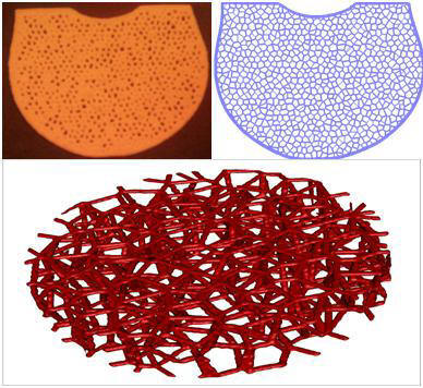
This research focuses on image-based multi-scale modeling, direct fabrication and mechanical analysis of bone and scaffold. Based on the digital images, the overall macroscopic geometry of bone can be acquired by traditional reverse engineering technology and the microscopic random trabecular network is described by a two-point correlation function and the function was then used to reconstruct the bone microstructure. It is shown that the reconstructed model is statistically equivalent to the original structure in the microscopic level. Biological tissue engineering design intention can also be integrated in the developed model. A voxel-based direct-fabrication process planning is developed and this makes the manufacture of complex tissue structure possible due to the elimination of CAD modeling and slicing process.

Stem Cell Printing: Cell Survivability and Possible Phenotype Reprogramming

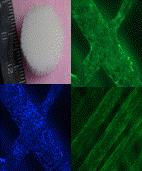
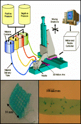
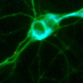
Cell printing is a powerful manufacturing tool to assemble biologics to bring structure and functional abilities to cell aggregates. Here we present a multi-nozzle bioprinter inspired by rapid prototyping technology. A CAD/CAM platform is integrated with solid freeform automation to assemble biologics in three-dimensional (3D) space. This system is used to execute micron-scale spatial control to repeatedly generate cell-laden constructs for scaffold guided tissue engineering applications; to include regenerative medicine, in vitro drug trials, and disease analogs. This is a physical process; which exposes the cells to exogenous mechanical stimulation. Cells, as a dynamic material, will fluidize and reorganize under mechanical stretch. Previous work has shown the pressurized micro-nozzle printing environment causes cell damage. This is well observed experimentally and the magnitude of the applied stress is analytically modeled. In this work we expand on existing characterization of cell response to include pluripotent mesenchymal stem cells (MSC). This cell was chosen for its self-regulating regenerative potential and non-immunogenic properties. The objective of this work is to observe the effect of printing on the MSC’s viability and phenotype. To accomplish this cells are recovered from the delivery matrix after printing under no stress (control), mid-range, and high stress conditions. Using fluorescent microscopy we visually label cell components. The conditions of these components were compared between experimental groups to determine the effect of printing. The metrics of comparison are the integrity of cell membrane, degree nuclear degradation, and morphology. Using this as evidence, we determine which cell features are affected and discuss ramifications to cell differentiation. We work backward from the experimental observations to describe the experience of the cell during printing. This knowledge can be applied during the selection of printing process parameters to consciously abstain from impacting cell behavior or potentially preferentially induce stimulation to direct cell behavior.

Bioprinting of 3-D Organ Chambers for Pharmacokinetic Study

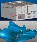
The conducted collaborative research between NASA-JSC and Drexel University was intended to explore the development and study of an in vitro three-dimensional Microfluidic Microanalytical Microorgan Device (3MD) for simulation of human response to drug administrations and toxic chemical exposure under microgravity and space environments. By fabricating a 3D in vitro tissue analog consisting of an array of channels with tissue-embedded chambers, one can selectively biomimic different mammalian tissues for a multitude of applications, foremost among them liver tissue for experimental study of drug metabolism and pharmaceutical screening of drug toxicity. The objective of research conducted at Drexel University was aimed at the achievement of high-throughput reproducible fabrication of bioprinted tissue constructs and 3D organ chambers, maintenance of structural integrity, the integration with a microfluidic platform, and the enhancement of cell viability with cell phenotype retention.

Modeling and direct fabrication of random heterogeneous tissue structures

This research focuses on image-based multi-scale modeling, direct fabrication and mechanical analysis of bone and scaffold. Based on the digital images, the overall macroscopic geometry of bone can be acquired by traditional reverse engineering technology and the microscopic random trabecular network is described by a two-point correlation function and the function was then used to reconstruct the bone microstructure. It is shown that the reconstructed model is statistically equivalent to the original structure in the microscopic level. Biological tissue engineering design intention can also be integrated in the developed model. A voxel-based direct-fabrication process planning is developed and this makes the manufacture of complex tissue structure possible due to the elimination of CAD modeling and slicing process.

Hybrid scaffold modeling and fabrication for tissue engineering application

Using multiple materials to freeform fabricate hybrid tissue scaffolds enable us to produce scaffolds with complex architectures to meet many needs of growing tissue. For instance, poly-caprolactone may be used for structural support while fibrin is used for cell attachment and alginate is used to provide a diffusion network for nutrient transport. The long-term goal of this research is to explore a feasibility of designing and fabricating scaffolds in which multiple tissues will be able grow within a single scaffold, restricted to regions for which they are intended by manipulation of materials and architecture.

Biomimetic design and fabrication of load bearing tissue scaffolds/replacements

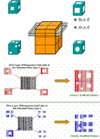
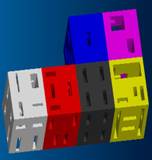
The design of 3D tissue scaffolds for tissue engineering application should, if possible, biomimic the complex hierarchy and structural heterogeneity of the replaced tissues. The objective of this research is to develop a computer aided tissue engineering approach for reconstruction, characterization, and the design of load bearing tissue scaffold informatics model. A biomimetic approach for modeling, design and fabrication of tissue scaffolds with intricate architecture, porosity and pore size is proposed. An Interior Architecture Design (IAD) approach which can be applied to generate scaffold layered freeform fabrication tool path without forming complicated 3D CAD scaffold models is developed. This IAD approach involves: applying the principle of layered manufacturing to determine the scaffold individual layered process planes and layered contour; defining the 2D characteristic patterns of the scaffold building blocks (unit cells) to form the interior scaffold pattern; and the generation of process tool path for freeform fabrication of scaffolds with the specified interior architectures.

Biopolymer deposition for freeform fabrication of tissue constructs

Polymeric scaffolds have been utilized in tissue engineering as a technique to confide the desired proliferation of seeded cells in vitro and in vivo into its architecturally porous three-dimensional structures. The ideal manufacturing of scaffolds may include cells simultaneously deposited along with the scaffolding materials, growth factor, and other nutritional and biological species. This research is attempted to fabricate biopolymer-based tissue scaffold at a bio-friendly environment, and develop a multi-nozzle biopolymer freeform deposition system. Studies on the biopolymer deposition-ability, 3D scaffold structural formability, and the construction of 3D hydrogel scaffold with living cells under different process parameters, such as nozzle sizes, types, regulating pressure and loading, the property of the biopolymer and the cross-linking agents are currently pursued, along with the study of the process-dependent cellular tissue engineering behavior of 3D tissue constructs.

Topological and transport connectivity for the tissue scaffolds

Scaffold design, porosity characteristics and the scaffold topological connectivity directly affect cell attachment, survival, proliferation, growth and guide new tissue formation. Cell survival and continued growth depend on delivery of nutrients and removal of waste. This dependence requires scaffold design to have pathways or connections allowing fluid and mass transport to cells throughout the scaffold. Collaborating with Computer Science researchers, this study establishes topological connectivity criteria, analyzes optimal transport architectures, and develops 3D skeleton and Earth Mover's Distance based algorithm for topological matching between designed tissue scaffolds to insure suitable connections for scaffold flow, mass transport, properties and fabrication.

Computer-aided tissue engineering approach for advanced tissue scaffold design

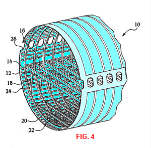
By using computer-aided design in conjunction with rapid prototyping and tissue engineering, computer-aided tissue engineering (CATE) has the power to explore many novel ideas that push the envelope of conventional scaffold designs by incorporating biomimetic and non-biomimetic features. CATE can be used to design and create scaffolds with controlled internal and external architecture; scaffolds with vascular channels of different sizes; modular scaffolds with interconnecting subunits; multi-layered scaffolds with spongy and compact regions; scaffolds with artificial structures such as chambers for drug delivery.

Polymer Extrusion using Precision Extrusion Deposition

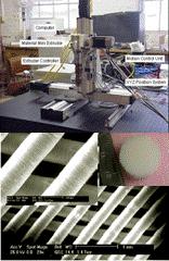
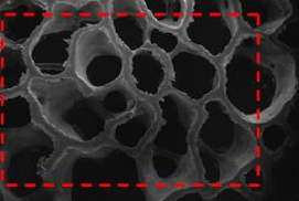
Successes in scaffold guided tissue engineering require scaffolds to have specific macroscopic geometries and internal architectures in order to provide the needed biological and biophysical functions. Freeform fabrication provides an effective process tool to manufacture many advanced scaffolds with designed properties. Using a novel Precision Extruding Deposition (PED) process technique, Poly-є-Caprolactone (PCL) scaffolds with a controlled pore size of 250 μm and designed structural orientations were fabricated. The scaffold morphology, internal micro-architecture and mechanical properties were evaluated using SEM, Micro-Computed Tomography (µ-CT) and the mechanical testing. Preliminary biological study was also conducted to investigate the cell responses to the as-fabricated tissue scaffolds. The results and the characterizations demonstrate the viability of the PED process to the scaffold fabrication as well as a good mechanical property, structural integrity, controlled pore size, pore interconnectivity, and the anticipated biological compatibility of the as-fabricated PCL scaffolds.

Computer-aided characterization for effective mechanical properties of porous tissue scaffolds

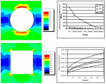
This research is attempted to develop a computer aided characterization approach to evaluate the effective mechanical properties of porous tissue scaffold. Process of computer-aided characterization and its interface with design model, development of a computational algorithm for finite element implementation and numerical solution of asymptotic homogenization theory is developed. Application of the algorithm to characterize the effective mechanical properties of porous Poly ε-Caprolactone scaffold manufactured by precision extruding freeform deposition, and a parametric study of the process and design parameter to the structural properties of tissue scaffold are conducted.

Image Guided Craniofacial Reconstructive Surgery

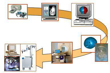
This is a collaborative research with Dr. Piatt, Chief of Neurosurgery at St Christopher’s Hospital for Children. Critical to the success of craniofacial surgery is the surgeon’s accurate perception of the anatomy of the deformity. This research is to develop a biomodeling and its application for quantitative control of craniofacial reconstructive procedures. Computed tomographic (CT) images of the patient’s skull are used to construct a 3D model of the deformity. Based on this model, a virtual reconstructive surgery is performed for both the deformed and reconstructed skulls and the physical medical prototypes are fabricated using a 3D Rapid Prototyping system. The reconstructed skull is then CT scanned. Employing the proprietary software of the BrainLab® surgical image guidance system, the virtually reconstructed image data set can be superimposed or “fused” with the original data set. The BrainLab® system then enables the surgeon to verify each step of the reconstruction procedure in real time, and discrepancies from the actual and the ideal can be corrected at the region of deviation.

Back to top

Research Collaborators

Dr. Mauli Agarwal, Department of Biomedical Engineering, University of Texas- San Antonio (biomaterials, tissue engineering)

Dr. Fred Allen, School of Biomedical Sciences and Engineering, Drexel University (cellular biology, tissue engineering)

Dr. Yuehuei An, Medical University of South Carolina (animal testing, in vivo study)

Dr. Dennis Blackmore, NJIT (algebraic algorithm, mathematical modeling)

Dr. Steve Gonda, NASA JSC (bioreactor, microgravity, tissue engineering)

Dr. Selçuk Güçeri, Dept of Mechanical Engineering, Drexel University (Fabrication of ceramic-ceramic composites, Fused deposition rapid prototyping of ceramics, nanotechnology)

Dr. Frank Ko, Dept. of Materials Science and Engineering, Drexel University (biomaterials, tissue engineering)

Dr. Alan Lau, Dept of Mechanical Engineering, Drexel University (computational analysis and simulation, fracture mechanics)

Dr Peter Lelkes, School of Biomedical Sciences and Engineering, Drexel University (Cellular tissue engineering, cellular biology)

Dr. Ming C Leu, University of Missouri, Rolla (CAD/CAM, virtual reality, manufacturing)

Dr. Michael Liebschner, Dept of Bioengineering, Rice University (bioengineering, biomechanics)

Dr. Feng Lin, Dept of Mechanical Engineering, Tsinghua University (CAD/CAE/CAM)

Dr. Joe Piatt, St. Christopher Children’s Hospital, Philadelphia (neurosurgery, Computer-Assisted Craniofacial Reconstructive Surgery)

Dr. William C. Regli, Dept of Math and Computer Science, Drexel University (Internet computing, artificial intelligence, geometric computation)

Dr. Caroline Schauer, Department of Materials Science and Engineering, Drexel University (biomaterials)

Dr. Ali Shokoufandeh, Dept of Computer Science, Drexel University (mathematic graph theory, pattern recognition, data clustering, gesture recognition)

Dr. Francis Wang, NIST (biopolymers)

Dr. Yongnian Yan, Dept of Mechanical Engineering (CAD/CAM, Tissue Engineering)

Back to top